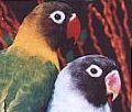
The Beak
The upper and lower jaws are covered by a hard keratinized structure known as the rhamphotheca. This horny material is continuously growing and worn down by the normal activity of the bird. The tip of the upper beak has a rich supply of blood and both the upper and lower beak have an abundant supply of nerve endings. Injury to the beak, especially the tip, can be extremely painful. Birds may be so uncomfortable that they refuse to eat.
The beak may become severly damaged as a result of an attack by a larger bird or other animal. Any cracks or fissures must be repaired to prevent food and water from invading the beak, leading to bacterial or fungal infection. Fortunately acryllics can be used to repair the damaged beak, enabling the bird to eat, groom, and move about until the tissue is replaces. The horny covering of the upper beak – the rhinotheca – is replaced in approximately six months while the Keratin of the lower beak – the gnatotheca is replaced in two to three months.
Congenital defects may occur, causing the beak to be abnormal.
Scissors Beak
Scissors beak is a condition in which the upper and lower beaks cross. Maindibular prognathium is a condition in which the lower jaw is longer and justs out, cathing the tip of the upper beak. This is especially common in cockatoos.
In the very early stages, corrective measures may be taken using wires and acrylics to correct these defects. After a certain stage, however, these become extremely difficult if not impossible to correct.
Overgrowth of the Upper Beak
Overgrowth of the rhinotheca (upper beak) may occur with liver disease. The beak usually has small bruises as well. This is most frequently seen in budgies on an all seed diet. Bloodwork, xrays, and a liver biopsy help support the diagnosis.
Rubber Bill
An extremely soft beak known as “rubber bill” may be seen in birds fed a diet low in calcium and Vitamin D. This is due to insufficient mineralization of the beak and is most commonly seen in doves and pigeons fed a wild bird seed diet without supplementation, instead of the proper colubiform diet.
Chronic Nasal Infection – Rhinitis
Chronic nasal infection or rhinitis can cause the development of longitudinal grooves in the upper beak as a result of the chronic discharge.
Knemidokoptes Mites
Knemidokoptes mites cause a disease known as “scaly face” or “scaly leg”. These mites are most common in budgies, but may occur in other types of birds as well. These burrowing mites cause proliferative lesions on the beak, legs, feet and cloaca. Although this mite is most common in young birds, it can affect adults also. Diagnosis is by the characteristic hyperkeratosis and scrapings of the skin. This disease can be treated by a drug that kills the mites. Left untreated, the beak will become severly disfigured and the bird may be unable to eat.
Beak and Feather Virus
The beak may develop fissures, cracks, and splits as well as overgrow as the result of beak and feather virus. The horny covering may even separate from the underlying bone and mucosa. This is extremely painful. Bacteria and fungi often invade the damaged beak. The bird is in a great deal of pain and often stops eating. This damage is progressive and very little can be done to make the bird more comfortable. Secondary infrections can be treated with antibiotics and antifundals, but the disease usually leads to the demise of the bird.
The Orapharynx
Tongue lacerations can occur as a result of fighting or gettin the tongue caught in the toy. These tongue lacerations require suturing because the tongue is in constant motion and will not heal if left alone.
Vitmain A Deficiency
Vitamin A deficiency is frequently seen in psittacine birds fed an all seed diet. The choanal papillae, (the small projections around the choanal opening in the the roof of the mouth) become blunted. The lining of the submandibular salivary glands undergoes a change known as squamous metaplasie. Swellings and excessive amounts of mucus are produced in the oral cavity. These swellings may occur on the roof of the mouth or under the tongue and contain a thick white material. These can interfere with eating. Treatment consists of lancing these, antibiotics and Vitamin A supplementation as well as improving the diet. (These changes also occur in the repiratory tract as well.
Internal Papillomatosis
Internal papillomatosis is an infectious disease of suspected viral etiology that affects new world parrots. Macaws, hawk-headed parrots, amazons and conures can be affected. Small wartlike growths known as papillomas can be found in the oral cavities, as well as in other places in the body. The papillomas are white or pink and have a cauliflour or cobblestone appearance. Common locations in the oral cavity are the base of the tongue, the margins of the choana and the glottis. These lesions may not be easy to see unless the beak is held open so that the oral cavity can be closely examined. If clinical signs do occur, they may include wheezing, salivation, difficulty swallowing, difficulty breathing and open-mouthed breathing. Bile duct and pancreatic duct carcinoma has been linked with internal papillomatosis.
Candidas
Candidas is an infection caused by the yeast Candida albicans. It can cause plaques and exudative lesions in the mouth which cause difficulty eating and drinking. Young birds – especially cockatiels – are most frequently affected. Prolonged antibiotic treatments, malnutrition, and concurrent illness can casue an adult bird to develop candida. This condition is treated with antifungal medication.

