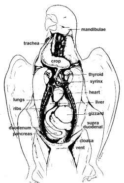
 Digestion in birds involves a lot of organs, each performing a specific function. It begins with entry of food via the beak and ends with the exiting of refuse at the vent. Food is broken down and absorbed for use along the way.
The Mouth
The avian beak or rostrum is composed of bone and an outer horney Keratin sheath or rhamphoteca. Keratin replacement continues throughout the life of the bird. In large parrots, the keration is worn down by digging, eating and chewing on hard objects and is completely replaced in 6 months on the upper bill and in 2 – 3 months on the lower bill. The upper beak is the maxillary rostrum and the lower beak is mandibular rostrum. The cutting edges of the beak are tomia, which take the place of teeth. In some parrots, the underside of the upper beak has ridges for holding food and maintaining the edge of the lower beak. The tip of the upper and lower beak is richly supplied with nerve endings.
Beaks replace the lips and teeth of mammals and vary in shape, size, length and function according to the type of diet consumed. Seed-crackers such as finches have a short conical beak, while birds of prey such as hawks have a powerful hooked beak for tearing flesh.
The choana is a median fissure in the palate which connects the oropharynx inside the mouth with the nasal cavity. Birds do not have a soft palate as mammals do. Numerous projections or papillae are found at the edge of the choana. These papillae often become blunted with a vitamin A deficiency.
A small slit like opening, the infundibular cleft is located behind the choana. This permits communication between the pharanyx and eustachean tubes of the ears. Abundant lymphoid tissue is found in the wall of this infundibular cleft.
The tongue, just as the beak, is adapted to the type of food the bird consumes. Woodpeckers have a long narrow protrudible tongue which functions as a spear – allowing them to extract insects. Parrots, birds of prey, and finches have short, thick, fleshy tongues which allow them to manipulate their food. Fowl and pellicans have nonprotrusible tongues with caudally-directed papillae which allow the food to be easily shoved to the back of the mouth for swallowing. Birds do not have an epiglottis to prevent food from entering their larynx. Instead, the opening of the glottis closes during swallowing.
Numerous small salivary glands are found in the oral cavity. These are best developed in birds that eat a dry diet such as seed and insect eaters, and least developed in birds that eat a moist diet such as fish. Mucus is the major substance secreted by the avian salivary glands. It acts as a lubricant in swallowing.
The epithelial lining of the mouth in some species – especially passerines (songbirds) – is brightly colored – – which stimulates the parents to feed the chick when it begs for food.
The Crop
The esophagus transports food from the mouth to the stomach. Many species of birds – such as parrots have an enlarged area of the esophagus known as a crop or ingluvies. Several types of crops exist. Gulls and penguins do not have a crop, while ducks, geese and song birds possess a small fustorm dilitation of their esophagus. The crop stores food temporarily and allows food to be softened before it enters the stomach. Pigeons and doves produce “crop milk” that they feed to their young for the first two weeks after hatching. Other species – such as parrots – will regurgitate food that has been stored and softened in their crops to their young.
The Stomach
Birds have a two part stomach, a glandular portion known as the proventriculus and a muscular portion known as the ventriculus or gizzard. Hydrochloric acid, mucus and a digestive enzyme, pepsin, are secreted by specialized cells in the proventriculus where chemical digestion begins. The gizzard has a thick muscular wall and plays an important part in the mechanical digestion by crushing and grinding food. The gizzard is lined by a cuticle or hardened carbohydrate and protein complex.
Fish and meateaters have a thin-walled sacklike stomach that is adapted for storage rather than mechanical digestion. The distinction between the proventiculus and the ventriculus is difficult to determine. Seedeaters (such as parrots), insectivores and herbivores have a very well developed gizzard adapted for mechanical breakdown of indigestible food. The proventiculus is easy to distinquish from the ventriculus in these birds.
The Intestines
The small intestine is the primary site of chemical digestion and nutrient absorption. It is divided into a duodenum, jejunum and ileum. These are arranged in u-shaped loops in the abdominal cavity. The large intestine is composed of paired caeca and a short rectum. The shape and size of the caeca vary in different species of birds. The caeca of the chickens are well developed while those of the passerines (song birds) are very small. Caeca are very rudimentary or absent in some species such as parrots and pigeons. Caeca contain lymphoid tissue. Cellulose breakdown by bacteria occurs in the caeca.
Chemical digestion and absorption of food occurs in the small intestine. Water resorption and temporary storage of waste material occurs in the cloaca and rectum.
Other Organs
The pancreas lies in the duodenal loop. The pancrease produces amylase, lipase and proteinases including trypsine. These are important enzymes for chemical digestion. It also produces the hormones insulin and glucogon which regulate blood sugar.
The large liver is divided into a right and left lobe. In most species the rught lobe is larger than the left. In some species – such as passerines – the right lobe is subdivided. The liver has numerous functions among which is the production of bile. Bile is important in the breakdown of fat. Bile is stored in the gallbladder.
Some species, such as parrots and pigeons, lack a gallbladder. In these species, bile is releaed directly into the small intestine.
The digestive tract is sterile at hatching. Altricial chicks – or chicks born blind and helpless – are innoculated with micrflora when parents feed them. Precocial chicks – chicks that are able to feed on their own shorly after hatching, are innoculated by bacteria from ther environment during feeding. In addition, a process known as “cloacal drinking” occurs in altricial birds by which microflora are “sucked” into the vent from the nest to colonize the posterior digestive tract.


Can you repair an esophageal trough on a parrot? If so can they talk after having it repaired?