One of nature’s true marvels is the design of the avian eye. In general, birds possess some of the best vision capabilities in the animal kingdom. Birds see in color which is important in recognizing food and danger and is possibly used in mating rituals. Birds also see with enormous accuracy and at long distances. Some have very advanced depth perception and motion detection capabilities, while others see well at night. A bird’s eyes are quite large in comparison to the size of its body and are extremely important to its survival.
Nocturnal birds such as owls make very efficient use of light. Other birds, have finely tuned focusing ability, enabling them to see accurately at long distances. In fact some birds of prey (such as raptors) have developed enhancements to the eye which enable them to focus at both far and near distances and to accurately judge the speed of moving objects.
The following examples show various ways that humans have attempted to use this superior ability.
After World War II, the military experimented with using birds to help locate flyers downed in the sea. The birds could see downed pilots at much greater distances than did the human observers.Companies that make pills (like asperin) or some candies, need final inspectors on the assembly line to remove the broken ones and to leave only perfect ones for packaging for the consumer. Human workers doing this type of work need frequent and long rest periods. A test using birds for this task proved that birds could work longer and be much more accurate.
Geese have been taught to eat the bad insects from crops while leaving the good insects alone. Their eye sight seems to be suited to this task.
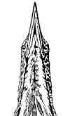
 The position of a bird’s eyes on its head varies by type of bird. Birds of prey have wider heads with the eyes set apart and facing forward, similar to humans. This gives them binocular vision or depth perception and enables them to judge the speed and distances of prey and other objects.

There are a number of diseases which can affect the eye, such as cataracts, conjuntivitis and absesses. Because birds rely so heavily on their eyesight, any problems should be treated immediately.
Although the anatomy of the avian eye is similar to that of mammals, a few reptilian characteristics remain. These reptilian characteristics are the pectin structure within the globe of the eye and the scleratic ring of bony plates – scleral ossicles that support the shape of the globe. Birds have enormous eyes relative to their body size and the keenest vision of all vertebrates.
The shape of avian eyes may be flat, globose, or tubular and varies according to the type of bird. “Flat” eyes are found in diurnal (awake during the day) birds with narrow heads such as doves, pigeons and parrots. They have a short anterior-posterior (front to back) axis, so that the image that falls on the retina is small, resulting in lower visual acuity. “Globose” (more rounded) eyes are found in birds with wider heads such as raptors and passerines (canaries, finches). The anterior-posterior axis is greater than in flat eyes, increasing visual acuity (focusing quality). “Tubular” eyes are characteristic of nocturnal birds of prey, such as owls.
The postion of the avian eye varies according to the shape of the skull. Pigeons, doves and parrots have narrow skulls with laterally placed eyes. Birds of prey, such as raptors, have broader skulls with more frontally positioned eyes. Laterally positioned eyes provide a large field of vision but a small binocular vision. Binocular visions means that both eyes are focused on the same object and move in a coordinated fashion.
Anatomy of the Eye
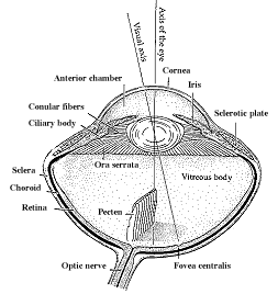
 The eye is composed of three “coats” or layers. The outer coat is the sclera. This is a strong white fibrous coat which maintains the shape of the eye, protects its internal structure, and serves as the attachment point for the eye muscles. The clear front portion of the eye is the cornea. Light passes through the cornea and into the pupil behind it. A series of small, overlapping bones – the scleral ossicles – are located in the sclera encircling the cornea. These bones strengthen the eyes and provide attachment for the ciliary muscles.
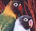
The middle coat or layer of the eye is the vascular tunic. It is composed of the iris, the ciliary body and the choroid. The iris is a thin sheet of muscle fibers and connective tissue that forms a diaphragm in front of the lens, controlling the amount of light entering the eye. The pupil is the circular open space at the center of the iris. The iris contains pigment that gives the eye its color. In some species of birds, the iris coloration changes with age or sex of the bird. For example, white and pink female cockatoos have a red iris, while males have a dark brown to black iris.
The ciliary body suspends the lens by zonular fibers. The ciliary body is located at the base of the iris. It suspends the lens and alters the shape of the lens, changing the focal point of the eye. Ciliary processes or folds formed by the ciliary body produce a thin fluid known as aqueous humor. This fluid is located between the cornea and the iris and between the iris and the lens. It maintains intraocular pressure and helps maintain the global shape of the eye. The ciliary muscles are very important for visual accomodation. The choroid is a thick, vascular, darkly pigmented layer that coats the retina and helps nourish both the sclera and retina.
The third coat or innermost layer of the eye is the retina. It is very sensitive to light and contains photo receptive rods and cones. Cones are important for visual acuity and color vision and are most numerous in nocturnal birds. The retina of birds is different from mammals in that it is thick and has no blood vessels. The pecten is a vascular, folded structure projecting from the avian retina. It is thought to aid the choroid layer in supplying the retina with nutrients and in transporting oxygen and carbon dioxide. The optic nerve centers the retina in the lower quadrant of the back of the eye.
The area of sharpest visual accuity of the retina is in the macular disk. The fovea centralis is a tiny pit in the center of the mocular disk. It is the area of maximal optical resolution. Most birds have single fovea. Birds that pursue fast moving prey – such as falcons – possess two foveate areas in each eye giving them very accurate perception of distance and speed.
The avian lens is softer than that of mammals – a very important feature in rapid visual accommodation. Ciliary muscles change the shape of the lens to focus on near or far objects, a process known as accommodation. Predatory birds such as owls have long, tubular eyes and cannot readily accommodate their eyes to focus on very near prey. They often have to move away from their prey to better see it.
The vitreous body is a clear, jelly like substance that fills the eye between the lens and the retina. The viscosity of the vitreous supports and maintains the shape of the eye.
A small moveable membrane, the nictictating membrane (or third eyelid) is located in the corner of the eye. This membrane moves across the eye, spreading tear secretion produced by the gland of the nictitating membrane and a lacrinal gland. This tear secretion moistens the cornea and protects against micro organisms.

